A 76 year old lady presents with low back, buttock, and posterior thigh pain. The pain is worse whenever she stands or walks and relieved with sitting or laying down. She rates the pain as 8 out of 10. She used to live an active, independent lifestyle but now she has difficulty doing basic tasks like grocery shopping, household chores, or yard work. She has tried medications, heat/ice, physical therapy, TENs units, and injections--all without lasting relief.
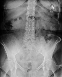 AP, lateral, and lateral flexion X-rays show degenerative disc disease at L4-5 and L5-S1, with grade 1 spondylolisthesis at L4-5.
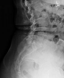 Preoperative lateral X-ray
 Preoperative flexion X-ray
 Axial T2 weighted MRI of the lumbar spine. L2-3 level is shown depicting relatively normal anatomy and no lumbar stenosis.
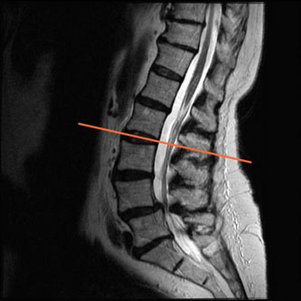 Sagittal T2 weighted MRI of the lumbar spine (line indicates location of L2-3 axial MRI image)
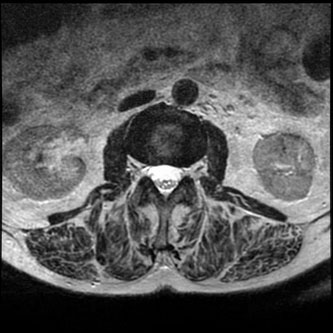
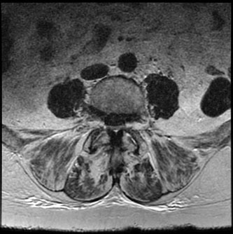 Axial T2 weighted MRI of the lumbar spine. L4-5 level has grade 1 spondylolisthesis and severe lumbar stenosis.
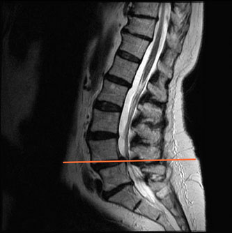 Sagittal T2 weighted MRI of the lumbar spine. (line indicates location of L4-5 axial MRI image)
The patient's x-rays showed spondylolisthesis at L4-5. Her MRI revealed severe lumbar stenosis at L4-5 as well.
Due to the severe stenosis and spondylolisthesis the patient agreed to proceed with a minimally invasive L4-5 decompression and transforaminal lumbar interbody fusion (TLIF).
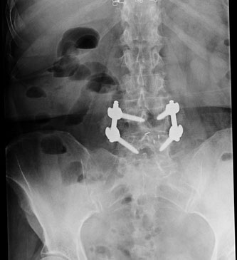 Postoperative AP X-ray shows interbody graft with screw and rod fixation at L4-5.
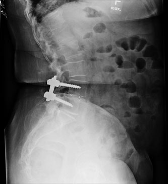 Postoperative lateral X-ray shows interbody graft with screw and rod fixation at L4-5.
After a short hospital stay the patient was discharged. At her two week follow up appointment her pain was gone and she was back to living independently!